
Implantation
Cleavage
Once syngamy has occurred in the ampulla of the uterine tube, a process termed cleavage begins. This refers to the mitotic divisions that occur in the first 3 days post-fertilisation. The first cleavage occurs around 30 hours after fertilisation and produces 2 daughter cells or blastomeres. These divide simultaneously at shorter and shorter time intervals producing double the number of blastomeres each time. By the time it reaches the uterus, which is around 72 hours after ovulation, there is a ball of 16 or more cells termed the morula. The morula is no larger than the zygote as cleavage produces smaller and smaller blastomeres. This gives an increase in the cell surface area to volume ratio allowing rapid nutrient uptake and waste removal.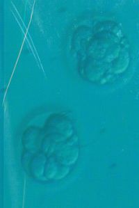
The morula migrates through the uterine tube and lies free in the uterine cavity for 4-5 days where it divides into 100 cells or so. Meanwhile the zona pellucida disintegrates and releases this structure, which is now called a blastocyst; a ball-like structure with a fluid-filled inner cavity (blastocoel). It is nourished by nutrients that were stored in the egg cytoplasm during oogenesis and by an endometrial secretion called uterine milk, which accumulates in a cavity. The blastocyst has;
- An outer layer of squamous (trophoectoderm) cells called the trophoblast: this develops into a variety of supporting structures including part of the placenta. It also plays an important role in the nourishment of the embryo.
- An inner cell mass called the embryoblast: this is destined to become the embryo itself.
Fig. 8. Blastocyst. Image courtesy of Flickr.
Implantation
Implantation of the blastocyst occurs in the endometrium of the upper posterior uterine wall of the fundus around 6/7 days after ovulation. The uterine cavity is narrow so there is more chance of attachment for the embryo.
Initial attachment may involve;
- Large carbohydrates: the embryo is floating in the uterine cavity and is trapped and slowed down by these carbohydrates – attach to receptors of the endometrium.
- Leukaemia Inhibitory Factor (LIF) is essential for implantation. If it is blocked no implantation can occur.
- Placenta Protein 14 (PP14 or Glycodelin): this is an immune suppressor which stops rejection. Recurrent miscarriages are often associated with a decrease in the levels of PP14.
The blastocyst proliferates rapidly as it meets the endometrium. Metaloproteases help burrow it into the endometrium and importantly the implanting blastocyst is not recognized as foreign. There is a ‘window of implantation’; the endometrium becomes hostile to the embryo after this. After implantation the endometrium becomes thicker and has a rich blood supply and the glands become more coiled and packed with glycogen; it becomes an energy rich sponge.
Trophoblast cells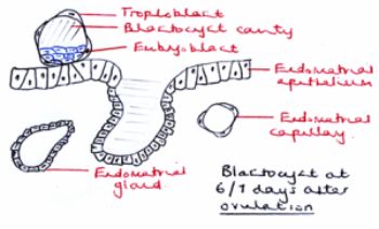 adjacent to the embryoblast secrete enzymes that stimulate thickening of the endometrium. It itself separates into 2 layers;
- The deep layer is called the cytotrophoblast – individual cells divided by membranes.
- The superficial layer is called syncytiotrophoblast – plasma membranes break down and the cells fuse into a multinucleate mass.
Fig. 9. Blastocyst 6/7 After Ovulation. Hand produced.
The latter grows into the uterus and digests endometrial cells along the way. The endometrium reacts by growing over the trophoblast and eventually enclosing it.
Implantation takes approximately a week and is completed by the time the next menstrual period would have occurred if the woman had not become pregnant.
Embryogenesis
During implantation the embryoblast undergoes embryogenesis, which is the arrangement of the blastomeres into 3 primary germ layers – ectoderm, mesoderm and endoderm. Embryogenesis begins with the formation of a narrow space called the amniotic cavity between the embryoblast and cytotrophoblast.
1. Embryoblast flattens into an embryonic disc composed of ectoderm and endoderm.
2. As the disc elongates a raised groove called the primitive streak forms along the midline of the ectoderm.
3. Cells on the surface migrate medially towards this groove, down into in and then laterally between the ectoderm and endoderm. This forms the middle germ layer, the mesoderm. The ectoderm and endoderm are epithelia composed of tightly joined cells. The mesoderm, however, is more loosely organised connective tissue called mesenchyme and its cells are more mobile.
4. At the end of embryogenesis the structure is an embryo – it is around 2mm long and 2 weeks old!