Overview of the Female Reproductive System
The male reproductive system is required to produce sperm, a function of the testes, and deliver them to the female tract through the penis. However, from here on it is the female reproductive system that is important; being the site of fertilisation right through to the expulsion of an infant at the end of pregnancy.
The component parts of the female reproductive system are;
The Ovary
The ovary is an almond-shaped organ nestled in the ovarian fossa, which is a depression in the dorsal pelvic wall. The female has 2 ovaries and they lie at either side of the pelvic cavity, held in place by connective tissue ligaments. They are the female gonads and so produce eggs as well as sex steroid hormones. The capsule of the ovary is termed the tunica albuginea and its interior is divided into 2 regions – the outer cortex which is where the germ cells develop and a central medulla occupied by the major arteries and veins.
The ovaries show asymmetry as usually only one is active each month, leading to the production of one egg. Hyperemia is observed in the active ovary; it swells and fills with blood becoming hotter and redder.
The internal genitalia are the uterine tubes, uterus and vagina, which form a pathway from the ovary to the outside of the body.

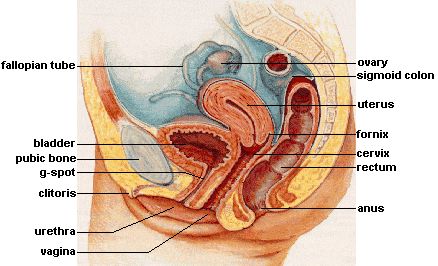
Fig. 2. Female Reproductive System (Lateral). Image courtesy of Wikimedia Commons.
Uterine Tubes
These are also called the fallopian or oviduct tubes and are the sites of fertilisation. They are canals about 10cms long from each ovary to the uterus. At their distal (ovarian) end they flare into an infundibulum that has multiple projections called fimbriae; these pick up the egg when it is ovulated.The middle part of the tube is the ampulla and near the uterus it forms a narrower isthmus. The uterine tube wall is predominantly composed of smooth muscle and its mucosa has an epithelium of ciliated cells and a smaller number of secretory cells. The cilia beat towards the uterus and, with the help of the muscular contractions of the tube, transport the egg in that direction.
The sperm travel up the tubes in order to meet the egg moving down through one of them from the ovary, its site of production. A fertilised egg embeds in the ampulla region and the tubes secrete various growth factors and glycoproteins for its early maintenance.
Uterus
The uterus is a thick muscular chamber that is suspended by a series of supporting ligaments. It opens into the roof of the vagina and usually tilts forward over the urinary bladder. It is the region of fetal growth, provides a source of nutrition and expels the fetus at the end of its development. It is a slightly pear-shaped structure; the superior broad portion is termed the fundus, the middle region is the body (corpus) and the inferior cylindrical region is the cervix.
Cervix
The uterus communicates with the vagina by a narrow passage through the cervix called the cervical canal. The superior opening of this canal into the body of the uterus is the internal os and the opening into the vagina is termed the external os. The canal contains cervical glands that secrete mucus; this is thought to prevent the spread of microorganisms from the vagina into the uterus. Close to ovulation the mucus becomes thinner allowing easier passage for sperm.
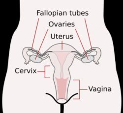
Fig. 3. Female Reproductive System (Front). Image courtesy of Wikimedia Commons.
Vagina
The vagina tilts dorsally between the urethra and rectum and is also termed the birth canal. It is around 10cms in length and is a single expandable tube with a lining composed mainly of muscle; the wall is thin but distensible. It allows the discharge of menstrual fluid, entry of the penis, admission of semen and the birth of a baby. The vagina has no glands but is lubricated by transudation (‘vaginal sweating’) of serous fluids through its walls and by the mucus of the cervical glands above it.
The female external genitalia include the clitoris, labia minora and labia majora.
